Today was treatment #24. I have four left to the full breast, and then 5 "boosters" to the scar area only. I googled to try to find out more about this last detail and this was the only relevant article:
Clips and scar as the guidelines for breast radiation boost after lumpectomy
F. Kovnera, f1, R. Agaya, O. Merimskya, J. Stadlerb, J. Klausnerc and M. Inbara a Tel-Aviv Sourasky Medical Center and Sackler Faculty of Medicine, Tel-Aviv University, Israelb Department of Surgery “A” Tel-Aviv University, Israelc Department of Surgery “B” Tel-Aviv University, Israel Accepted 12 April 1999. Available online 12 May 2002.
Abstract Background and Aims: Breast-conserving therapy in early breast cancer is equally effective as mastectomy, with advantages of cosmesis and quality of life over mastectomy. Local control is improved when entire breast irradiation is combined with a radiation boost to the tumour bed.
Methods: Localization of the tumour bed was compared in 45 consecutive patients using surgical scar and radiopaque clips placed intra-operatively in the lumpectomy cavity.Results : The area (A) of the radiation boost field and volume (V) of the tumour bed, designed on the basis of scar (AS and VS), were 1.4 times larger than those designed on the basis of the clips (AC and VC). AS and VS missed about one-quarter of the tumour bed which had been delineated by clips intra-operatively, while about one-half of it encompassed tissues beyond the AC and VC.
Conclusions: A boost planned by scar dimensions can miss a substantial portion of the tumour bed, compromising local control. On the other hand, a substantial amount of breast tissue beyond the tumour bed can be unnecessarily irradiated, compromising cosmetic treatment results. Thus, the scar provides an inadequate landmark for radiation boost planning in breast-conserving therapy.
So, I am not quite sure what to think. Probably a lot has changed since this article was published in 2002. What they did today was to apply a colored plastic patch in the shape of my scar to the area on my breast where the scar is; a crescent shaped scar that runs from approximately 1 o'clock to 4 o'clock. Lots more Sharpie lines. I think this tech really wanted to be a tattoo artist. Then Dr. P was called and finally came in to check the placement. For this treatment, the accelerator head has a kind of focused beam coming through a lens with a diameter of about 5-6", and it comes pretty close to my skin.
Tuesday is meet with the Dr. day. I wait an interminably long period of time for Dr. P. Everything is good (except my blood pressure!).
Tuesday, January 29, 2008
Interview and Job Status
I had a job interview on Friday, which seemed to go well. I have yet to hear anything. I'm worried because when I was looking for a job in 2005 - 2006, I had four or five interviews before I had a job offer.This client had asked for me by name, so unless I said something that put her off I am hopeful.
If I lose my job, I will finish my treatments (Just 9 left!) and go on unemployment while I look for something new. It doesn't appeal to me right now, but maybe it is an opportunity.
If I lose my job, I will finish my treatments (Just 9 left!) and go on unemployment while I look for something new. It doesn't appeal to me right now, but maybe it is an opportunity.
Sunday, January 20, 2008
The Comments
I made changes to allow anonymous comments, but I will moderate them. Please leave a comment, even if you are just passing through, to say hello and let me know you were here.
Your position has been eliminated!
Friday afternoon I learned that the agency that I work for as a contractor to the federal government had had it's budget cut by 1.2 million. Apparently that translates into 13 contractor positions, and that includes me. There are two positions currently open which I am qualified for, and hopefully they will like me and offer the position to me. My current job ends January 31, or at treatment 26. This being middle aged, having cancer, trying to keep your job stuff sure is no fun. Anyone who tells you the economy is doing great is lying to you.
Radiation tattoos and markups
This first image shows the markups right after my first few treatments.

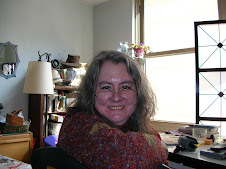
This image shows my markups at treatment #18. You can see the radiation line going across my chest and under my arm. The scar from the lymphectomy is included in the radiation from under neath on my left side.
 These markings are used by the radiology techs to line you up in the machine just so. They use laser beams in a darkened room to make sure you are just right. Thursday, I lay down on the table, put my arm up in the sling, and scooted up. Perfect! It was actually a little creepy if you think about it. So I was somewhat relieved on Friday when we had to do the usual poke and slide around routine to get me in position.

This image shows my markups at treatment #18. You can see the radiation line going across my chest and under my arm. The scar from the lymphectomy is included in the radiation from under neath on my left side.

Healing Shawl

I got a phone call a few days ago from a woman from my church (http://www.rruuc.org/) who is part of a caring circle of Mindful Knitters. They have made me a healing shawl. This is the Unitarian Universalist version of a prayer shawl. Cynthia brought it to me last night. She also had her 3 yo son with her so we didn’t get to converse too much. It is crocheted in muted earth tones, clay, silvery grey. In the natural sunlight that floods my apartment in the afternoon, I see it is rose and a deeper rose.
It came with this blessing
May grace be upon this shawl…
Warming, comforting,
Enfolding and embracing.
May this mantle be a safe haven…
A sacred place of security and well-being…
Sustaining and embracing in good times as well as difficult ones.
May the one who receives this shawl
Be cradled in hope, kept in joy, graced with peace, and wrapped in love.
Blessed Be
Adapted from Prayer of Blessing for a Completed Shawl by Janet Bristow.
Unfortunately, one of the side effects of the radiation treatment is my skin has turned pink like a mild sunburn. I am having trouble staying cool, not warm! But comforting, yes. The Alpha cat loves it already. He thinks it was made for him.
Also note that since my surgery, this 10 year old cat sits on the couch with me, next to me, for the first time ever.
Wednesday, January 9, 2008

I was googling around, hoping I might be able to steal another woman’s experiences with radiation. It seems each persons experience is so different and unique, so I am stuck with writing up my own.
On December 14th, I met my mom at the Brookland/Catholic University Metro stop to keep my appointment with Dr. Porrazo.
He is my radiation oncologist and today is the day I will be whirled around in the CT scanner and marked up for my radiation treatments. You lie on a table about the size and comfort of an ironing board. The physicist, who introduces himself as Francis (IIRC, and another technician named Muhammad) has me lie down and then using the images on the CT scanner he marks my chest with a Sharpie. They have also taped some wires down to my chest. Since I have my head turned to the right the whole time, I can’t really see what’s going on. I end up with an X in my cleavage, at my collar bone, just above my belly button, on either side at my waist… I’ll have to go look in a mirror to remember where they all are, but in total there are six. When they are satisfied with the positioning, they use a small pen shaped devise to permanently tattoo me in six places. I can scarcely feel this at all, but the one on my right side waist tickles when he tries to place it and I squiggle. The X’s are all covered with a round piece of plastic tape to preserve them for the actual treatment. Unfortunately, the one in my cleavage keeps melting, especially over the weekend. I am scolded over this; it’s the primary reference point. So I am stuffing cotton balls and tissue in my bra at night, and trying to sleep more on my back than my side.
I’ve described the SIM experience in my January 1st entry.
Today was my 11th treatment. Today actually went very well. Tuesdays are my day to meet with the doctor. Since I’ve been in treatment two weeks, they want to do blood work, primarily to check iron, hemoglobin, hemocrit. I ask if I can put it off til Wednesday, and get the go ahead. This works out well because I can get to WHC by 8:30 am, trip up to the lab, get poked and get downstairs for treatment. I was actually changing when one of the techs came back to get me, so I got in and out early. They have complained that they have back to back patients in 10 minute slots from 7 am to 12 noon. Unfortunately, many patients do not come on time, and things get backed up.
My primary complaint is that my skin is turning bright red like a minor sunburn. I am using Alba non-petroleum jelly all over my breast. I’ve also figured out that I spare myself discomfort by wearing a lighter weight turtle neck, especially in this warm weather we’ve been having. I check with the doctor to see if it is okay to take ibuprofen for the discomfort (yes) and melatonin for sleep (yes).
I have been waking up at odd hours, and then am not able to go back to sleep. Part of it is the anxiety of knowing I have to get up and get out of the house on time to catch the bus to get the WHC. Monday night I woke up around 2 am. I was able to go back to sleep, and dreamed an old flame had come to my bedroom and was making love to me. That was nice. I’m not sure what any lover would make of my Sharpie marked-up boob.
Friday, January 4, 2008
eight down
Well, I made it through my first week but not quite how I wanted. Today was treatment #8. My appointment is supposed to be at 8:50 am but most days they are not calling me until around 9:20 am. This morning I did not push it too much to get out the door early, and I was there by 8:50 am. Come to find that my treatment machine, the Alpha machine, is "down." I ask about this, it turns out the machine goes through a calibration routine and if it does not pass all the tests it does not come up. A good thing, I suppose. The first computers I worked on had this too, it was called IPL or Initial Program Load, and if it failed you were SOL. I was done and out to catch the bus by 9:45 and at work by 10:15 am. I'm going to put in a shorter day and take an hour of leave and call it a week.
My breast is starting to feel tender and pink already. I commented on this and was told by Mustaphe that dark people get darker and people "like me" get pink. It is "an expected side effect." I suppose eventually I will have a tan left breast. I had anticipated this but not so soon. Eight down, 20 to go of the "general" radiation series.
Dianne
Tuesday, January 1, 2008
Where I am now
I'm going to fast forward to get up to date. On Friday, Dec 21 I had my "simulation."
There is a good description of the procedures that the radiation physicist performed along
with the CT Scan, which I had done 12/14
http://wwwbooble.blogspot.com/2007/11/i-was-so-wired-today.html
I will try to update you with my version of this twirling and tattooing voodoo medicine
My mother came with me and waiting anxiously and alone in the waiting room. Then she got to meet Dr. Porrazo
He was very reassuring to her, explaining that the
radiation treatment is primarily prophalytic in my case, since most of my tumor
was intraductal.
Dr. Porrazo and the physicists come up with a treatment protocal by examining the images from the CT Scans. The next Friday, I show up for my "sim" and meet the radiologists. However, they cannot find my treatment protocal. This has been typical at WHC, it seems they have
too many computer systems and not much integration. My appointment is at 11:00 and at 11:20 they tell me the physicists are "still working" on my treatment plan.
Thanks god they did not need to take my blood pressure, because I am sure it is somewhere close to the roofline. Finally, they walk me into the treatment area, and position me according to the angles and degrees that are on the computer screen. By this time, however, I am so
tense that when they leave the room and check me on the computer, I am off my
"marks". All three of them come running back in, kind of like Keystone
cops, and rearrange me and try again. The team are taking X-rays to make
sure that they have interpreted the instructions correctly. Finally it is done.
The treatment team on the Alpha machine are Mustapha, Ade
(two very dark men from Africa) and Roselia.
My first treatment is 12/26, and it goes much like my
"sim." Lots of running back and forth and re-arranging me. Isn't
"try to relax" an oxymoron? Treatment 2 is a bummer because they get to me
20 minutes late, but I got my zaps on the first try. Treatment 3 goes
smooth as silk, I get my zaps all the first time, and catch the 9:05 bus and get
to work by 9:30. So this is doable. Three down, 30 to
go.
I'm getting radiated!
Here's a good photo of the linear accelerator:
http://www.whitememorial.com/content/services/cancer/index.asp
And here: Click on the photo to see an
enlargement.
http://news-service.stanford.edu/news/2007/april18/med-accelerator-041807.html
Think horses not zebras

The wisdom is to think horses not zebras when your hear hoofbeats. In my case, it is a zorse. The biopsy shows cancerous cells and the diagnosis is Ductal Carcinoma in situ September 26, 2007. This is considered Stage 0 cancer. When Dr. B does the surgery, the biopsy of the tumor finds that 1% is invasive. I will have to have radiation. The tumor is also tested to see if it is estrogen progesterone sensitive. It is, and I will have to take an anti-hormone, probably tamoxifen.
The equipment

You lie on the table with your boob hanging down, the doctor sits at the computer, and the tech tries to get you lined up correctly. You get some anesthesia locally, and they shoot you with the needle. It's not too bad. This is the same setup they use the day of surgery to wire me. I'm still trying to figure that out!
Stereo tactic biopsy
Dr. Butler did not think my mammo looked all that "suspicious" but he said he would not overrule the KP radiologist. so the next step was to get a biopsy,
in my case a stereotactic breast biopsy.
http://www.radiologyinfo.org/en/info.cfm?pg=breastbixr&bhcp=1
There's lots of info on line about the procedure, but what I really wanted was a picture of the table. There is a good one in the booklet they gave me, so I may try to get that
scanned in.
What does the equipment look like?
A specially designed table is used for stereotactic biopsy. The patient is lying face-down with her breast projecting through a hole in the table. The actual biopsy is done below the table after raising it to gain access to her breast. The procedure also may be done with the patient upright in a chair. An upright study may be best for those women who might have difficulty climbing onto the table or who are unable to lie prone for any reason. You must not move during the procedure.
A paddle-shaped instrument compresses the breast during biopsy. (Like a mammogram) A tray is nearby containing all of the equipment necessary for the biopsy.
Actually in my case the computer was across the room. The procedure was done by a nurse and a doctor. There were two places of concern, and they tried very hard
to get lined up for one punch, but it never happened. What should have taken about 5 minutes took over 30 minutes and by then I was very cranky.
In addition to the specialized equipment needed for x-ray-guided breast biopsy, specially trained technologists and physicians perform the procedure. The images are obtained not with x-ray-exposed film as in conventional mammography, but using computerized or digital imaging in place of a film cassette. This reduces x-ray exposure to the breast and also permits the images to be viewed on a computer monitor seconds after exposure—compared with the several minutes needed to develop x-ray film. The principle of stereotactic biopsy is that a lesion can be located precisely in three dimensions by calculating its apparent change in position on angled x-ray images. The first x-ray locates the abnormality in the breast, after which two stereo views are obtained, each angled 15 degrees to either side of the initial image. The physician then marks the lesion electronically on the stereo images. The computer calculates how much the lesion's position appears to have changed on each of the stereo views, and in this way is able to determine its exact site in three-dimensional space.
The biopsy instrument used in this procedure is called a vacuum-assisted device (VAD), which consists of an inner needle with a trough extending from it at one end and an overlying sheath. When the sheath is retracted, a vacuum is used to pull breast tissue into the needle trough. The outer sheath rapidly moves forward to cut the tissue and collect it in the trough.
An advantage of the VAD is that the needle is inserted only once into the breast without having to withdraw the needle after each sampling. Biopsies are obtained in an orderly manner by rotating the probe, assuring that the entire region of interest will be sampled.
The first step is to clean the skin and inject a local anesthetic. A small nick is made in the skin and the tip of the biopsy needle is advanced to the previously calculated site of the lesion. At this point stereo images are again obtained to confirm that the needle tip is actually within the lesion. Usually six to 12 samples are collected when the VAD is used. Then a final set of images is obtained. If they show that the lesion has been mostly or completely removed, a small clip is left at the biopsy site so that it can be easily located if the lesion proves to be cancer. Once the biopsy is complete the skin opening is covered with a dressing; it need not be sutured. You will be told to avoid strenuous activity for 24 hours after returning home, but then usually will be able to resume normal activities

Meet the Surgeon
I called Kaiser to get an appointment with a surgeon.
People complain that with an HMO you don't get to pick your doctor. I really don't
need the stress of making any more decisions. KP refers me to Dr. John
Butler.
His main office is at the Washington Hospital Center, but in
September he was also working at the Capitol Hill Kaiser office. This is a
convenient walk for me from my office at BLS.
He refers me for a stereotactic biopsy at the Kaiser
facility in Kensington. He assures me that 85% of the time, this is
benign.
Subscribe to:
Posts (Atom)